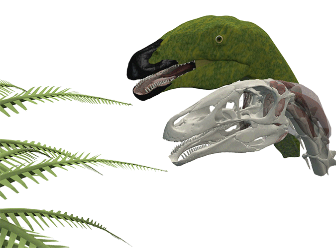
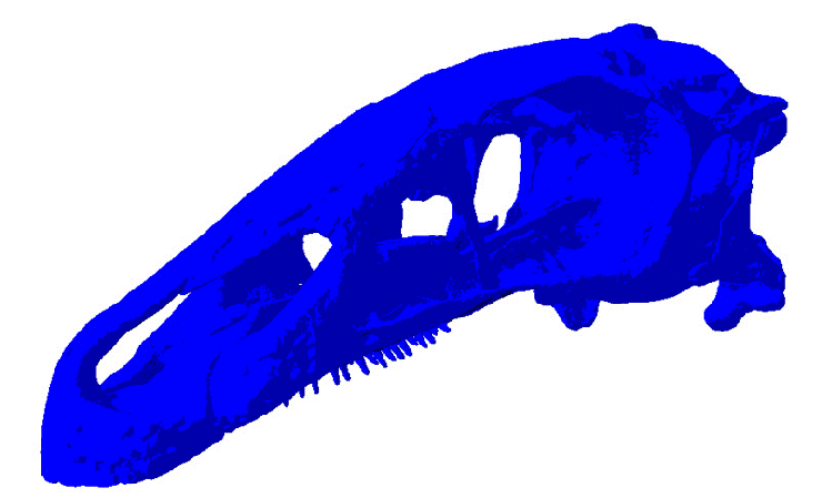
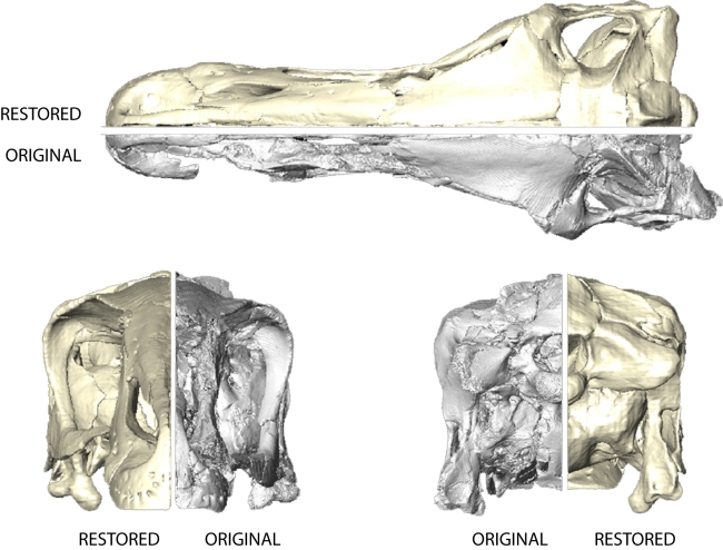
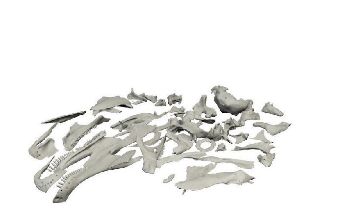
DIGITAL RESTORATION OF FOSSILS
By their very nature, fossils are often incompletely preserved, distorted and deformed, when there are found. Millions of years of fossilisation processes have left their mark on the fossils. But also during excavation, collection and preparation fossils can further be damaged. However, this can present a serious problem for the study of extinct organisms. For example, knowledge on taxonomic relationships of fossil taxa relies on a large part on the (preserved) form. But also information about the appearance, the behaviour and the ecology of extinct species depends on detailed knowledge of their anatomy.
My research focuses on using different digital techniques to restore the original form of fossils. The restored three-dimensional models can then subsequently be used to reconstruct relevant soft-tissue structures and ultimately permit further investigation of function, such as feeding or locomotion.
More information and examples can be found here:
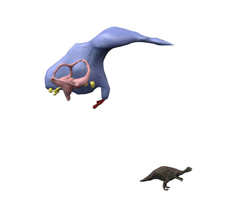
DIGITAL RECONSTRUCTION OF FOSSIL SOFT TISSUES
Fossils usually consist of preserved hard parts such as bones and teeth in vertebrates and mineralised shells in invertebrates. In contrast, soft tissues are only rarely preserved in the fossil record, yet detailed knowledge of soft-tissue structures is paramount to understanding the palaeobiology of extinct organisms. However, novel computational techniques, including CT scanning and digital visualisation, provide versatile tools to reconstruct soft-tissues, such as the brain anatomy and the musculature, of fossils virtually:
Palaeoneurology, the study of the anatomyof the brain, inner ear and nerves in fossils, holds a large potential to learn more about the evolution of the brain in extinct animals. For my research, I am interested how based on the morphology of these soft tissues, sensorial capabilities (i.e. hearing, smelling, vision) in fossils can be studied.
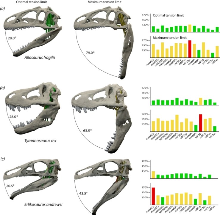
Similarly, the musculature plays a fundamental role in an animal’s life. For example, the jaw muscles are primarily associated with feeding and strongly influence feeding processes, diet and the ability to occupy specific ecological niches. In order to gain insight into functional properties, such as muscle forces and bite forces, jaw movement or possible gape angle, the musculature has to be reconstructed. Digital reconstruction methods and virtual models offer powerful tools to do this.
More information and examples can be found here:
FUNCTIONAL MORPHOLOGY AND BIOMECHANICAL MODELLING
The field of functional morphology analyses the relationship between anatomical form and function and behaviour. In fossil organisms, function is often difficult to reconstruct. However, by using a range of biomechanical modelling techniques, such as Finite Element Analysis (FEA) or Multibody Dynamics Analysis (MDA), coupled with CT scanning and digital visualisation, it is possible to investigate the form/function-relation of extinct animals. These techniques are particularly powerful tools to not only compare different skeletal morphologies, but also to test hypothetical models and different behavioural scenarios.